Radiology Sections - Abdominal Imaging and Cross-sectional Interventional Radiology
Contact:
Raquel Alencar, MD
617-732-7724
In this section, radiography, fluoroscopy (real-time x-rays), CT, and MRI are used to diagnose diseases within the abdomen and pelvis. Additionally, a number of procedures (primarily biopsies and drainage of abscesses) are performed here using CT and/or ultrasound guidance. The “standard” abdomen and pelvis CT scan consists of a single scan through the abdomen and pelvis following the administration of IV (if the patient is a suitable candidate) and oral contrast and is commonly used to evaluate abdominal pain (appendicitis, diverticulitis, pancreatitis, etc.), fever (abscess), trauma (solid or hollow viscous injury), and patients with a history of malignancy (query metastases). A number of additional special protocols have been developed for specific clinical situations, and some of these include the renal stone protocol, the CT urogram, the adrenal washout CT, CT colonography, and others, which you will learn more about during your time in the reading room. Abdominal and pelvic MRI protocols are generally tailored to the clinical question, but specific organs commonly evaluated for malignancy and other disease processes include the pancreas, liver, kidneys, and prostate. Abdominal radiographs (KUB for kidneys, ureters, bladder) and fluoroscopic studies are interpreted in a nearby but separate location. Radiographs are most often obtained to evaluate for signs of bowel obstruction or perforated viscous, whereas fluoroscopy involves the use of oral contrast (most commonly barium) to image the lumens of various organ systems (most commonly the GI tract). Though fluoroscopy has become less frequently used since the advent of cross-sectional imaging, these studies can still provide valuable information that no other modality can offer.
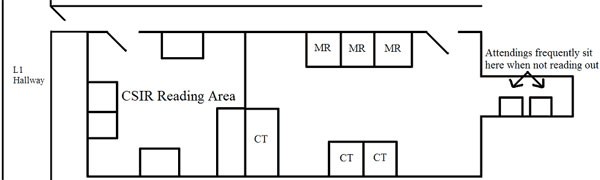
The main abdominal CT/MRI reading room is located on L1 near the Abram’s conference room. The abdominal radiograph/fluoroscopy reading room is also located on L1 and is situated down the hall from the CT/MRI reading room and across the hall from ultrasound (see BWH L1 map). It is recommended that you split your observation time between these two areas.