Radiology Sections - Neurologic Imaging
Contact:
Eileen Walsh
617-732-7260
In this section, CT and MRI are almost exclusively used to diagnose diseases of the brain, spine, and extra-cranial head and neck. Radiography has a very limited role in this area. Noncontrast head CT is an integral step in the initial workup of presumed ischemic stroke to evaluate for hemorrhage in patients being considered for thrombolytic therapy. It is also the preferred test for initial radiologic evaluation of patients with traumatic head injury, sudden onset severe headache, and other patients in whom acute intracranial hemorrhage is suspected. The CT angiogram is commonly used in the acute setting to evaluate for intracranial aneurysm as well as carotid or vertebral dissection. To answer almost all other clinical questions related to the brain, MRI is the preferred modality. Other studies you will see interpreted within this section include temporal bone CT, spine MRI and CT, and CT/MRI of the neck, face, and sinuses.
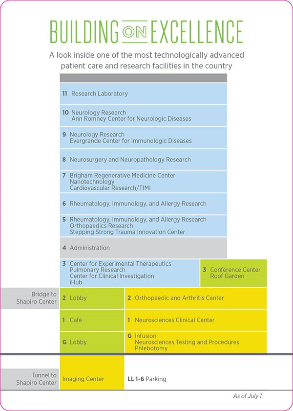
The neuroradiology reading room is located on LL-1 in the new Building for Transformative Care.
During your time in this section, you should observe the interpretation of at least one brain CT, one brain MRI, one spine CT, one spine MRI, and one perfusion study. You should also attempt to observe at least two of the following procedures: lumbar puncture, myelogram, CT-guided head and neck procedure, and catheter angiogram (again, ask the fellow to review the clinical history, imaging, and indication for the procedure ahead of time).