Radiology Sections - Thoracic Radiology
Contact:
Laura Freeman
617-732-6285
In addition to the ubiquitous chest radiograph, CT and MRI images of the chest are also interpreted within this section. Chest CT is commonly used to diagnose and to follow pneumonia, lung nodules, lung tumors, mediastinal and hilar masses, pleural diseases including empyema and mesothelioma, and interstitial lung disease. Different protocols are available for use in different situations. Pulmonary embolus CT’s are performed to detect pulmonary embolism and are also interpreted within this section, and some ultrasound-guided (when the lesion is peripheral) and CT-guided procedures (mainly lung biopsies) are also performed here. Thoracic PET/CT’s are also interpreted by this section, though the actual readout occurs in the nuclear medicine area. You should ask to be present for at least one of these readouts (see goals below).
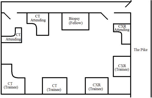
The chest reading room is located directly off the Pike (see BWH Pike map).
During your time in this section, you should observe the performance of at least one lung biopsy. Introduce yourself to the thoracic imaging fellow, who will be stationed at the biopsy or “procedure” desk. Ask the fellow to review the patient’s history, imaging findings, and the indication for the biopsy with you before going to observe the procedure.